- Home
- Resources
- Patient Release Form to obtain your records from other Physician
- Patient Release Form to release your records to other Physician
- Insurance and Billing
- What should I bring to Appointments
- Glossary in English
- Glosarios de Examenes de diagnostico
- High School Sports Injuries
- Stretching Exercises for Sports
- Childhood Obesity and bone, joint & muscle health
- News
- Staff
- Conditions
- Galleries
- FAQ
- Zink Blog
- Contact
 Call Us : 1-407-894-0088
 joan@zinkmd.com
Knee Injuries ACL
Knee Injuries ACL
Recognizing an ACL injury
People who play basketball, volleyball, soccer, or football, or who ski are most likely to injure their ACLs when they slow down, pivot or land after a jump.
If you injure yours, you may not feel any pain immediately. You might hear a popping noise and feel your knee give out from under you.
Within a few hours, you’ll notice swelling at the knee. The knee will often hurt when you try to stand on it. It’s important to keep weight off the knee until you can see your health care provider, or you may injure the knee cartilage. You should use an ice pack to reduce swelling and keep the leg elevated. If needed, use a pain reliever. If you must walk, use crutches and be sure to see a doctor right away to have your knee evaluated.
Prevention & Risk Assessment
Tips for Preventing an ACL Knee Ligament Injury
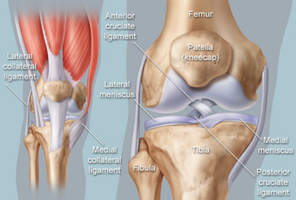
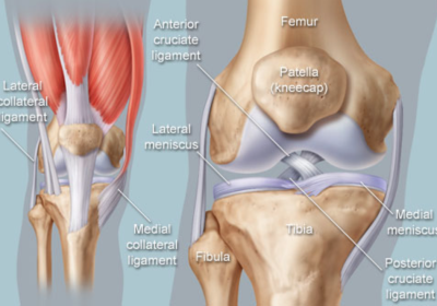
The knee is a joint where 3 main bones join: the femur, or thigh bone; the tibia, or shin bone; and the patella, or knee cap. Several ligaments attach to the femur and tibia and give the joint strength and stability. One of these, the anterior cruciate ligament (ACL), is in the center of the knee and limits rotation and the forward movement of the tibia.
The ACL is most often stretched or torn by a sudden twisting motion — when, for example, your feet are planted 1 way and your knees are turned another. You can also injure your ACL by quickly changing the direction in which you’re moving; by putting the brakes on too quickly when running; or, when landing from a jump. A woman’s body structure and hormones cause more force on the ligaments, increasing the likelihood of injury during sports and athletic activities.
Prevention
Many ACL injuries can be prevented if the muscles that surround the knees are strong and flexible.
Prevention focuses on proper nerve and muscle control of the knee. Exercises aim to increase muscle power, balance, and improve core strength and stability.
The following training tips can reduce the risk of an ACL injury:
- Train and condition year round.
- Practice proper landing technique after jumps. This involves bending your knees to absorb the force and keeping them in line with your feet.
- When you pivot, crouch and bend at the knees and hips. This reduces stress on the ACL.
- Strengthen your hamstring and quadriceps muscles. The hamstring muscle is at the back of the thigh; the quadriceps muscle is at the front. The muscles work together to bend or straighten the leg. Strengthening both muscles can better protect the leg against knee injuries.
Knee Conditions
- Chondromalacia patella (also called patellofemoral syndrome): Irritation of the cartilage on the underside of the kneecap (patella), causing knee pain. This is a common cause of knee pain in young people.
- Knee osteoarthritis: Osteoarthritis is the most common form of arthritis, and often affects the knees. Caused by aging and wear and tear of cartilage, osteoarthritis symptoms may include knee pain, stiffness, and swelling.
- Knee effusion: Fluid buildup inside the knee, usually from inflammation. Any form of arthritis or injury may cause a knee effusion.
- Meniscal tear: Damage to a meniscus, the cartilage that cushions the knee, often occurs with twisting the knee. Large tears may cause the knee to lock.
- ACL (anterior cruciate ligament) strain or tear: The ACL is responsible for a large part of the knee’s stability. An ACL tear often leads to the knee “giving out,” and may require surgical repair.
- PCL (posterior cruciate ligament) strain or tear: PCL tears can cause pain, swelling, and knee instability. These injuries are less common than ACL tears, and physical therapy (rather than surgery) is usually the best option.
- MCL (medial collateral ligament) strain or tear: This injury may cause pain and possible instability to the inner side of the knee.
- Patellar subluxation: The kneecap slides abnormally or dislocates along the thigh bone during activity. Knee pain around the kneecap results.
- Patellar tendonitis: Inflammation of the tendon connecting the kneecap (patella) to the shin bone. This occurs mostly in athletes from repeated jumping.
- Knee bursitis: Pain, swelling, and warmth in any of the bursae of the knee. Bursitis often occurs from overuse or injury.
- Baker’s cyst: Collection of fluid in the back of the knee. Baker’s cysts usually develop from a persistent effusion as in conditions such as arthritis.
- Rheumatoid arthritis: An autoimmune condition that can cause arthritis in any joint, including the knees. If untreated, rheumatoid arthritis can cause permanent joint damage.
- Gout: A form of arthritis caused by buildup of uric acid crystals in a joint. The knees may be affected, causing episodes of severe pain and swelling.
- Pseudogout: A form of arthritis similar to gout, caused by calcium pyrophosphate crystals depositing in the knee or other joints.
- Septic arthritis: Bacterial infection inside the knee can cause inflammation, pain, swelling, and difficulty moving the knee. Although uncommon, septic arthritis is a serious condition that usually gets worse quickly without treatment.
Knee Tests
- Physical examination: By examining the location of knee pain and looking for swelling or abnormal movement, a doctor gathers information about potential causes of damage or stress on the knee.
- Drawer test: With the knee bent, a doctor can pull (anterior drawer test) and push (posterior drawer test) the lower leg while holding the foot stable to check the stability of the ACL and PCL knee ligaments.
- Valgus stress test: Pushing the calf outward while holding the thigh stable, a doctor can check for injury to the medial collateral ligament (MCL). Pushing the calf inward (varus stress test), a doctor can look for injury to the lateral collateral ligament (LCL).
- Knee X-ray: A plain X-ray film of the knee is typically the best initial imaging test for most knee conditions.
- Magnetic resonance imaging (MRI scan): Using high-energy magnetic waves, an MRI scanner creates highly detailed images of the knee and leg. An MRI scan is the most-often used method of detecting ligament and meniscal injuries.
- Arthrocentesis of the knee (joint aspiration): A needle is inserted into the joint space inside the knee, and fluid is drawn out. Various forms of arthritis may be diagnosed through knee arthrocentesis.
- Arthroscopy: A surgical procedure that allows examination of the knee with an endoscope.
Related Portfolio
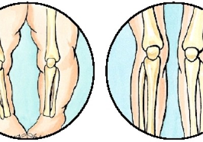
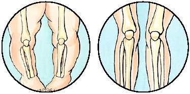
Pediatric Bowlegs
Pediatric Bowlegs What Are Bowlegs? Bowlegs are a condition in which the legs curve outward at an extreme angle at the knees while the child’s feet are together. Babies are born
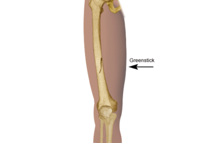
Broken Bones
Pediatric Broken Bones What is a fracture? A fracture is a partial or complete break in the bone. When a fracture occurs, it is classified as either open or closed: Open fracture
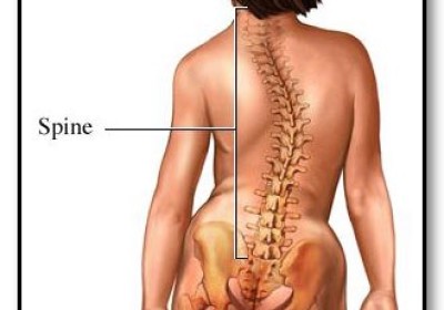

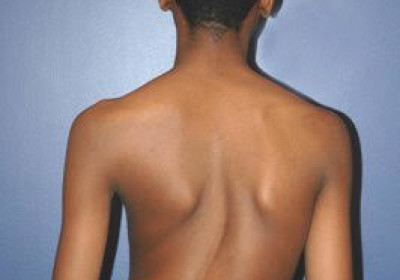

Pediatric Congenital Spine Deformities
Pediatric Congenital Spine Deformities What Are Congenital Spine Deformities? Congenital spine deformities are disorders of the spine that develop in a child before birth. The
Knee Injuries ACL
Knee Injuries ACL Recognizing an ACL injury People who play basketball, volleyball, soccer, or football, or who ski are most likely to injure their ACLs when they slow down, pivot